Shedding light on exceedingly rare biological events.
Autonomous Microscopy for Rare Cancer Biology
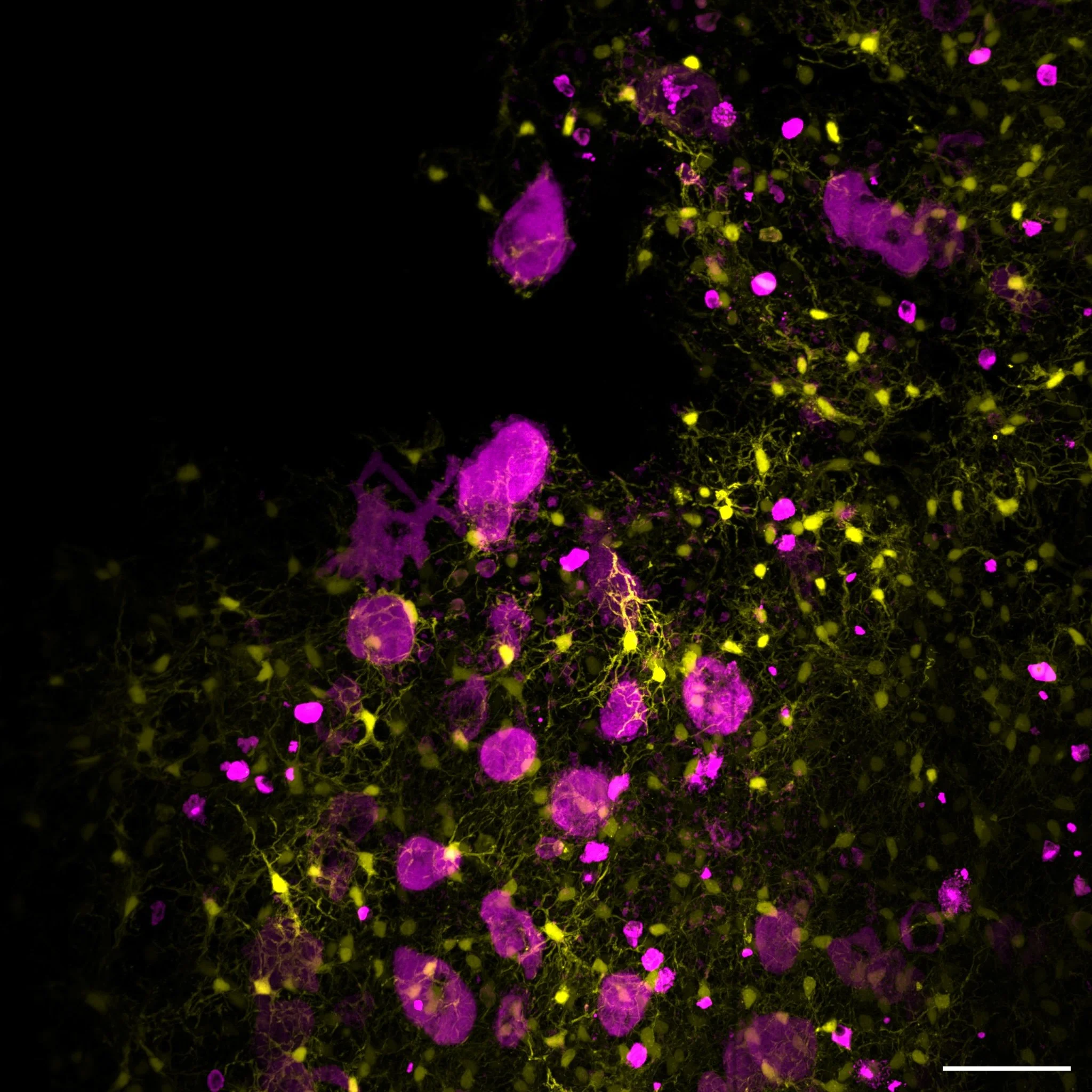
The Dean Lab develops open-source microscopes, adaptive acquisition software, molecular multiplexing workflows, and computational image-analysis tools to study rare biological events in intact tissues.
We are especially interested in how cancer cells survive, migrate, and colonize distant organs. These processes are difficult to observe because they are sparse, spatially complex, and often distributed across large tissue volumes. To make them measurable, we build imaging systems and computational workflows that can survey large specimens, identify biologically relevant events, and extract quantitative information across molecular, cellular, and tissue scales.
What We Build
Autonomous Microscopy
We develop self-driving microscopes that combine light-sheet imaging, real-time image analysis, and adaptive acquisition. These systems are designed to search large specimens, recognize relevant biological features, and automatically collect higher-resolution or higher-content data where it matters most.
Molecular Multiplexing
We develop optical, chemical, and computational workflows for increasing the molecular information content of fluorescence microscopy. Our approaches include spectral imaging, cyclic multiplexing, expansion microscopy, and integrated analysis routines for spatially resolved tissue measurements.
Open-Source Imaging Tools
We develop and disseminate open-source software, hardware, documentation, and analysis workflows for the microscopy community. Our goal is to make advanced imaging technologies easier to reproduce, adapt, and apply across biological systems.
Featured Tools and Resources
navigate
navigate is open-source Python software for microscope control, adaptive acquisition, and reproducible light-sheet imaging workflows. It is designed for both biologists using advanced microscopes and developers building new imaging systems.
Altair
Altair is an open-source light-sheet microscopy platform designed to make high-performance volumetric imaging more accessible to biological research labs.
Clearex
We develop computational routines for preprocessing, segmentation, object detection, feature extraction, spatial analysis, tracking, visualization, and large-scale microscopy data handling.
Funding
Our work is supported by collaborative programs in cancer imaging, biomedical technology development, microscopy dissemination, and quantitative cell biology. These projects support the development of autonomous microscopy, light-sheet imaging, molecular multiplexing, image analysis, and technologies for studying metastatic tumor formation in intact tissues.